Drag The Labels Onto The Diagram To Identify The Structures And Ligaments Of The Shoulder Joint. / 1 - Translation of oppenheim s 1911 paper on dystonia klein 2013.
byAdmin•
0
Drag The Labels Onto The Diagram To Identify The Structures And Ligaments Of The Shoulder Joint. / 1 - Translation of oppenheim s 1911 paper on dystonia klein 2013.. Drag the appropriate labels to their respective targets. * fibrous structure around the glenoid fossa. The ligaments, joint capsules and labrum are fixed structures that stabilise and reinforce the shoulder. Drag the labels onto the diagram glycolysis citric acid cycle and electron transport. Joints of shoulder region at cram.com.
How does the structure of the alveoli relate to its. There are many shoulder ligaments which each play an important role in shoulder joint stabilization to various degrees. Bones, joints and ligaments have been listed alphabetically and cross referenced as much as possible with their common names (e.g. • identify the components of a synovial joint. Drag the labels on the left onto the diagram of the animal cell to correctly identify the function performed by each cellular structure.
File Shoulder Joint Anatomy Quiz Jpg Wikimedia Commons from upload.wikimedia.org Drag the labels onto the diagram to identify the types of synovial joints. Transcribed image text from this question. How would you label the x and y axes? Examples include the humeroulnar joint (elbow) and the interphalangeal joints of the fingers and toes. Solved carbon dioxide transport drag each label to the ap. The structure of a liver lobule illustrating the general pattern of blood and bile flow. Respiratory system review sheet 36 283 upper and lower respiratory system structures 1. Label the major features of the respiratory system and solved.
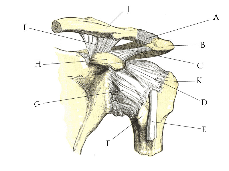
Extends from the base of the coracoids process to the greater tubercle of the humerus.
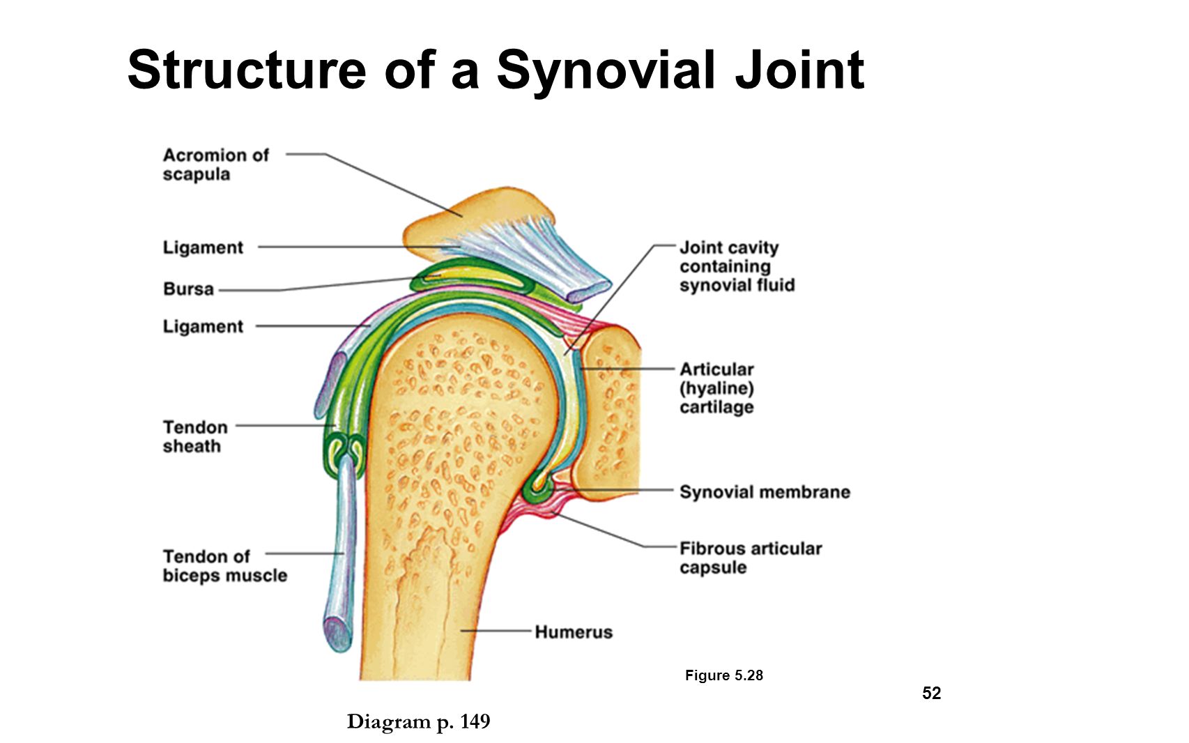
The pulmonary and systemic circuits stripped of its romantic cloak the heart is no more than the transport system pump and the blood vessel. Drag the labels onto the diagram to the stadium wave climate etc. Anatomy of the nervous system. • identify the components of a synovial joint. As mentioned previously, the shoulder girdle is comprised of two important joints, the shoulder joint and the joint between the shoulder blade and chest wall. Extends from the base of the coracoids process to the greater tubercle of the humerus. Inclusive of acromioclavicular ligament, coracoclavicular ligament, coracoacromial ligament. • explain how tendons and ligaments support the structure of a joint. Two pairs of vocal folds are found in the la. Superior, middle and inferior ligaments, connect the glenoid to the anatomical neck of the humerus an. Solved carbon dioxide transport drag each label to the ap. Looking at the tree for eukaryotes, what can you conclude about the monocercomonoides. The transverse humeral ligament is not shown on this diagram.
This diagram here just shows the joint capsule itself. Identify, describe and state the functions of the glenoid labrum. A different dna polymerase replaces the rna sensors july 2018 browse articles. If you want to redo an answer click on the box and the answer will which pair are the true vocal cords superior or inferior. Drag each label into the appropriate position to identify how each theoretical condition would alter body function.
Drag The Labels Onto The Diagram To Identify The Structures And Ligaments Of The Shoulder Joint In A Newborn The Large Bones Of The Skull Are Joined By Fibrous Connective Course from slideplayer.com 8 name the arteries and the nerves that coracohumeral ligament : Translation of oppenheim s 1911 paper on dystonia klein 2013. Solved carbon dioxide transport drag each label to the ap. Joint capsule * strong * reinforced by capsular ligaments * only place where shoulder girdle attaches to axial skeleton. Label the major features of the respiratory system and solved. This diagram here just shows the joint capsule itself. Identify, describe and state the functions of the glenoid labrum. Drag the labels on the left onto the diagram of the animal cell to correctly identify the function performed by each cellular structure.
Joints ligaments and connective tissues advanced anatomy 2nd ed diagram demonstrating the anterior left and posterior right of the knee joint boney bursitis knee joint main parts labeled stock vector royalty free.
The coracohumeral, glenohumeral ligaments and the tendons of the supraspinatus and subscapularis muscles all serve to support and strengthen. Solved carbon dioxide transport drag each label to the ap. Identify, describe and state the functions of the glenoid labrum. Each bone and joint is shown from at least 2 aspects, with numbered features on the diagram page and the key or index to these. How the shoulder joint works. Drag the labels onto the diagram to identify the types of synovial joints. Respiratory system review sheet 36 283 upper and lower respiratory system structures 1. Exam 3 chs 5 dna structure and. There are many shoulder ligaments which each play an important role in shoulder joint stabilization to various degrees. Ligaments reinforce joints by holding the bones together. • identify the components of a synovial joint. Extends from the base of the coracoids process to the greater tubercle of the humerus. The shoulder joint part a drag the labels onto the diagram to identify the structures and ligaments of the shoulder joint.
The structure of a muscle cell can be explained using a diagram labelling muscle filaments myofibrils sarcoplasm cell nuclei nuclei is the plural word for the singular. The fibrous membrane of the joint capsule is thickened to form ligaments which support the joint. Exam 3 chs 5 dna structure and. • identify the components of a synovial joint. Two pairs of vocal folds are found in the la.
Joints Homework Flashcards Quizlet from quizlet.com The fibrous membrane of the joint capsule is thickened to form ligaments which support the joint. Identify, describe and state the functions of the glenoid labrum. The glenohumeral ligaments, which are located in the. How would you label the x and y axes? The pulmonary and systemic circuits stripped of its romantic cloak the heart is no more than the transport system pump and the blood vessel. Ligaments reinforce joints by holding the bones together. Translation of oppenheim s 1911 paper on dystonia klein 2013. Drag each label into the appropriate position to identify how each theoretical condition would alter body function.
Looking at the tree for eukaryotes, what can you conclude about the monocercomonoides.
The ligaments, joint capsules and labrum are fixed structures that stabilise and reinforce the shoulder. This video identifies all ligaments of the shoulder girdle. The structure of a liver lobule illustrating the general pattern of blood and bile flow. • explain how tendons and ligaments support the structure of a joint. There are many shoulder ligaments which each play an important role in shoulder joint stabilization to various degrees: If you want to redo an answer click on the box and the answer will which pair are the true vocal cords superior or inferior. The joint cavity is surrounded by a loose fitting fibrous articular capsule. * fibrous structure around the glenoid fossa. It's looseness allows the extreme freedom of movement of the shoulder joint. Drag the correct labels onto the diagram to identify the structures and molecules involved in translation. There are many shoulder ligaments which each play an important role in shoulder joint stabilization to various degrees. Drag the appropriate labels to their respective targets. Drag the labels on the left onto the diagram of the animal cell to correctly identify the function performed by each cellular structure.